What Is an MRI? How It Works, What It Shows, and What to Expect
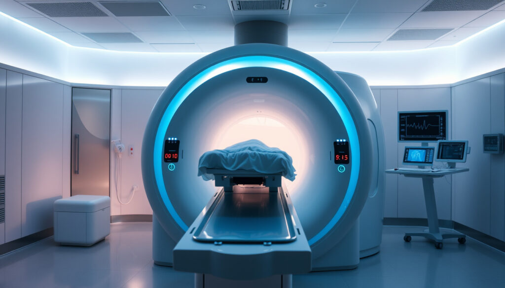
If you’ve ever been told you need to hold still inside a large, noisy machine for a while, you probably already know that MRI can feel a bit intimidating. Behind the loud knocks and the tunnel-like bore is a remarkably sophisticated technology that sees inside your body without a single X-ray. This guide covers exactly what an MRI is, how it works, how to prepare, and what sets it apart from other imaging methods.
Full name: Magnetic Resonance Imaging ·
Radiation used: None (uses magnetic fields and radio waves) ·
Typical magnet strength: 1.5–3 Tesla (clinical) ·
Scan duration range: 15–90 minutes ·
Developed: 1970s (first human scan in 1977) ·
Common uses: Brain, spine, joints, soft tissues, tumor detection
Quick snapshot
- MRI is a noninvasive imaging test that uses magnetic fields and radio waves (Johns Hopkins Medicine)
- No ionizing radiation is used (Mayo Clinic)
- Provides outstanding soft-tissue contrast (Johns Hopkins Medicine)
- Long‑term biological effects of repeated exposure to static magnetic fields are not fully understood but are considered negligible (Mayo Clinic)
- Optimal protocols for reducing patient anxiety during MRI are still being developed (Mayo Clinic)
- The safety of MRI in pregnancy during the first trimester is not fully established, and scans are avoided unless necessary (Mayo Clinic)
- First developed in the 1970s; first human scan performed in 1977
- MRI is increasingly used for disease detection, diagnosis, and treatment monitoring (National Institute of Biomedical Imaging and Bioengineering)
Six key specs give you the full picture in one glance.
| Full Name | Magnetic Resonance Imaging |
|---|---|
| Invented | 1970s; first human scan in 1977 |
| Radiation | None |
| Typical Magnet Strength | 1.5–3 Tesla (clinical machines) |
| Average Scan Time | 30–60 minutes (range 15–90) |
| Common Uses | Brain, spine, joints, abdomen, pelvis, blood vessels |
What exactly does an MRI show?
Common conditions diagnosed with MRI
- MRI produces detailed images of soft tissues, organs, blood vessels, and other internal structures (Johns Hopkins Medicine).
- It can reveal abnormalities such as tumors, inflammation, nerve damage, and joint issues (Johns Hopkins Medicine).
- MRI does not use ionizing radiation (Mayo Clinic).
- Bones and air‑filled spaces (e.g., lungs) appear dark or are not well visualized (Johns Hopkins Medicine).
Where CT excels at bone and acute bleeding, MRI owns the soft‑tissue domain — the difference drives every clinical referral.
The implication: if your doctor needs to see a torn ligament, a brain tumor, or damage to a disc, MRI is often the first choice.
Parts of the body typically imaged
- Brain and spinal cord
- Joints (knee, shoulder, hip)
- Heart and blood vessels (with contrast)
- Abdomen and pelvis (liver, kidneys, uterus)
What should you not do before an MRI?
Items to remove before entering the MRI room
- Remove all metal objects: jewelry, watches, coins, keys, glasses, hairpins, and removable dental work (Johns Hopkins Medicine).
- Leave credit cards and any magnetic stripe cards outside (UH Cleveland).
- You will typically be asked to change into a hospital gown because metal fasteners, zippers, buttons, and fabric fibers can interfere with the magnetic field (UH Cleveland).
Medical implant screening checklist
- Inform your doctor about any implants, pacemakers, cochlear implants, aneurysm clips, or metal fragments (UH Cleveland).
- Fasting is not always required, but for some abdominal MRIs you may be asked to avoid food/drink for 4–6 hours (Mayo Clinic).
- Tell the team if you have ever worked with metal or had an eye injury — an X‑ray may be ordered to check for metal debris (UH Cleveland).
Why this matters: The magnet is always on. Ferromagnetic objects can become dangerous projectiles, and certain implants can heat up or malfunction. The screening is not red tape — it’s a safety net.
The pattern: screening is the only layer between a routine scan and a preventable accident — always disclose everything.
How long will an MRI take?
Factors that affect scan time
- Most individual scans last 15–45 minutes; a full exam including setup may take up to 90 minutes (Johns Hopkins Medicine).
- The exact time depends on the body part being imaged and the number of sequences required (Johns Hopkins Medicine).
- Contrast injections add time for the injection and subsequent scanning (Mayo Clinic).
- You must lie still during the scan to avoid blurring (Johns Hopkins Medicine).
What to expect during the scan
- You lie on a sliding table that moves into the bore of the machine (Mayo Clinic).
- You will hear loud knocking or humming sounds — earplugs or headphones are provided (Johns Hopkins Medicine).
- You can communicate with the technologist via intercom throughout the exam.
The catch: the longer you have to stay still, the more anxious some people get. Knowing the timing upfront can help you mentally prepare.
What’s the difference between an MRI and a CT scan?
How the two technologies work
CT (computed tomography) uses X‑rays (ionizing radiation); MRI uses a strong magnet and radio waves (Mayo Clinic).
When each is preferred
Three contrasts, one pattern: each tool has its own sweet spot.
| Aspect | MRI | CT |
|---|---|---|
| Radiation | None | Uses X‑rays (ionizing) |
| Best for | Soft tissues (brain, muscles, organs) | Bones, lungs, acute bleeding |
| Speed | 15–90 minutes | Usually under 5 minutes |
The trade-off: if you need a rapid stroke assessment or a look at a fracture, CT wins on speed. But for a torn ligament or a subtle brain lesion, MRI’s soft‑tissue resolution is unmatched (Johns Hopkins Medicine).
Do I wear my own clothes in an MRI?
Why street clothes are usually not allowed
- Metal fasteners, zippers, buttons, and fabric fibers can interfere with the magnetic field (UH Cleveland).
- Some facilities allow scrubs or MRI‑safe clothing if it has no metal (UH Cleveland).
- Comfortable clothing without metal (e.g., cotton sweatpants and a t‑shirt) may be acceptable – check with your imaging center (UH Cleveland).
What to wear instead
- You will typically be asked to change into a hospital gown (Johns Hopkins Medicine).
- Remove all jewelry, watches, and any clothing with metallic threads or snaps (Johns Hopkins Medicine).
- Leave everything including keys, coins, and phone in a secure locker.
Patients who show up in metal‑free, comfortable clothes often skip the gown altogether — saving time and staying comfortable.
The implication: what you wear determines whether you breeze through or waste time changing — check ahead.
Steps to prepare for an MRI
- Remove all metal objects – including jewelry, glasses, hearing aids, and removable dental work (Johns Hopkins Medicine).
- Check with your doctor about implants – pacemakers, clips, coils, or any metal inside your body (UH Cleveland).
- Ask about fasting – for abdominal MRIs you may need to avoid food/drink for 4–6 hours (Mayo Clinic).
- Wear or bring metal‑free clothing – cotton sweatpants and a t‑shirt are often acceptable.
- Arrive early – allow time for screening, changing, and any contrast‑related questions.
For patients who follow these steps, the process flows smoothly. The only surprise left is the noise — and that’s what earplugs are for.
Clarity check: what we know and what remains open
Confirmed facts
- MRI does not use ionizing radiation.
- MRI is safe for patients without ferromagnetic implants.
- MRI provides superior soft‑tissue contrast compared to CT.
- Patients must remove all metal objects before entering the scan room.
What’s unclear
- Long‑term effects of repeated exposure to strong static magnetic fields are not fully understood but are considered negligible.
- The exact mechanism of how MRI affects certain tissues at the molecular level is still an active area of research.
- Optimal protocols for reducing patient anxiety during MRI are still being developed.
- The safety of MRI in pregnancy during the first trimester is not fully established, and scans are avoided unless necessary.
The pattern: confirmed facts are well-supported; the unclear items highlight where evidence is still evolving.
Perspectives from leading medical organizations
“MRI is a non‑invasive imaging technology that produces three‑dimensional detailed anatomical images. It is often used for disease detection, diagnosis, and treatment monitoring.”
National Institute of Biomedical Imaging and Bioengineering
“An MRI (magnetic resonance imaging) is a test that creates clear images of structures inside your body using a large magnet, radio waves and a computer.”
Cleveland Clinic
“MRI scans can be used to find out what’s causing any symptoms you’ve been having, check your bones and organs, and see how well a treatment is working.”
NHS (UK)
Three authoritative voices, one consistent message: MRI is a powerful, radiation‑free tool for seeing inside the body — and its role in medicine is only expanding.
lonestarneurology.net, touchstoneimaging.com, swdic.com, ncbi.nlm.nih.gov, youtube.com
For a detailed breakdown of how MRI differs from computed tomography, see this CT scan vs MRI comparison.
Frequently asked questions
Can I have an MRI if I am pregnant?
MRI is generally avoided during the first trimester unless the benefit clearly outweighs the risk. Always inform your doctor and the MRI team if you are or might be pregnant.
What is the difference between an open MRI and a closed MRI?
Open MRI machines have a wider opening and are less confining, making them suitable for patients with claustrophobia. However, they often have lower magnet strength (typically 0.3–0.7 Tesla) compared to closed 1.5–3 Tesla units.
What does an MRI scan feel like?
You will not feel the magnetic field or radio waves. You may feel a slight vibration and hear loud tapping sounds. The table may move slowly as the machine positions you.
Can I eat or drink before an MRI?
For most MRIs you can eat normally. Some abdominal scans (e.g., MRCP) require fasting for 4–6 hours. Check with your imaging center.
How soon will I get the results of my MRI?
A radiologist interprets the images and sends a report to your referring doctor. Results are usually available within a few days.
Is it safe to have multiple MRI scans?
Yes, because no ionizing radiation is used. MRI is considered safe for repeated use, though you should always discuss the medical necessity with your doctor.
What should I do if I am claustrophobic during an MRI?
Talk to your doctor beforehand. They may prescribe a mild sedative. Some facilities offer open MRI or use mirrors and music to reduce anxiety.
For anyone who has ever wondered what an MRI scan actually involves, the answer is straightforward: it is a quiet revolution in medical imaging that has already saved countless lives. Whether you are a patient scheduling a first scan or a healthcare professional referring a case, the choice between MRI and other modalities rests on one question — do you need to see the soft tissue clearly? If yes, the magnetic field holds the answer.
More related posts
 Swollen Lymph Nodes Neck: Red Flags and When to Worry
 Civil War (2024) Film: Controversy, Box Office, and Review
 Where Do Bed Bugs Come From? Origins & How to Get Rid of Them
 How to Remove Oil Stains from Clothes (Even After Drying)
 FC Barcelona Players 2025-26: Full Roster & Squad Analysis
 Tomodachi Life: Living the Dream – Price, Release, Switch
 Mueller, She Wrote Twitter: Account, Controversy & Influence
 St. Louis Cardinals vs Phillies Match Player Stats: May 14